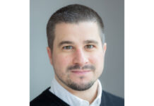
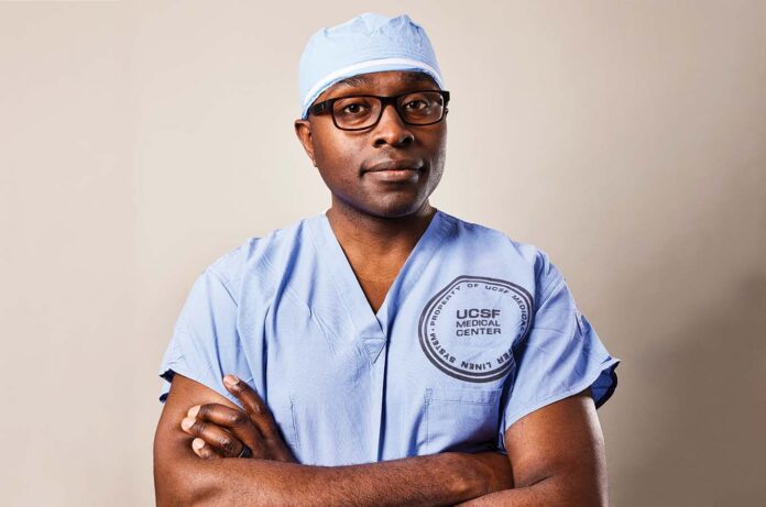
On a clear day in October, Shawn Hervey-Jumper, MD, rises before dawn at his home in the Oakland hills, a one-time horse ranch that he shares with his wife, two daughters, and a menagerie of animals, including a dog, three goats, and two kittens named Pepper and Cobbler, after his grandmother’s peach cobbler. He packs a raspberry yogurt and a mug of black tea. Then he heads to the Helen Diller Medical Center at UC San Francisco, where he will attempt to excise a deadly tumor from a conscious human brain.
Hervey-Jumper specializes in awake brain surgery, in which patients are alert and engaged during parts of the procedure in order to prevent severe brain damage. His patient this morning, Gina, is 31 years old. It’s not her first time under Hervey-Jumper’s knife. Six years ago, after she had a seizure following a car accident, doctors discovered a mass the size of a plum in the top left lobe of her brain, near the areas that regulate movement and speech. The tumor had been spreading slowly, but there was no cure. Without surgery, she was told, she might still have several years left to live; an operation could double that time, or maybe more. Hervey-Jumper had removed the original mass, but like crabgrass, the cancer sprouted back around the hole.
Now, in the operating room, Gina (whose name has been changed to protect her privacy) rests inside a cave of blue surgical drapes. She lies on her right side, facing the cave’s opening, her skull secured with a clamp. An anesthesiologist perches before her, monitoring her vital signs. At her back, on the other side of the drapes, technicians and research assistants lay out instruments and tend to various machines. Standing at the crown of Gina’s head, Hervey-Jumper peers down through a window in the drapes onto a shaved patch on her scalp. With a gloved hand, he touches a scalpel to her pale skin.
For the moment, Gina sleeps. Over the years, Hervey-Jumper has gotten to know her well. He knows that she is close to her mother and sister and has been helping to raise a young niece. Her vibrant personality reminds him of his grandfather, who was full of love and life – someone he liked to “kick around with.”
Hervey-Jumper lived with his grandparents, Perry and Charlotte Hervey, for the first years of his life. Perry, a custodial worker who planted a peach tree in their yard, was diagnosed with pancreatic cancer when Hervey-Jumper was 4. What scared him more than his grandfather’s impending death was the way the disease diminished him. “He lost a bit of himself,” Hervey-Jumper says. He hopes to spare Gina from such an experience for as long as he can. If this surgery goes well, he believes she will see her niece grow up.
He slices through her numbed scalp smoothly, swiftly, assuredly. The blade cuts an arc that follows the earlier incision. Then he peels back the flesh to expose dull, beige bone. “We’re going to drill,” he tells the room.
Meanwhile, as her sedation is turned off, Gina begins to gradually wake up.
How does a brain surgeon start his day? By feeding his goats.
Extraordinary Feats
At age 43, Hervey-Jumper is the latest in a long line of UCSF neuro-surgeons known for removing tumors once deemed inoperable. A soft-spoken man with broad shoulders and a round, youthful face, he grew up in rural Massachusetts, in a town surrounded by cranberry bogs. After he was born, his mother enrolled in nursing school, and he often tagged along with her to clinics and seminars, which kindled his interest in medicine.
“Early on, I thought I wanted to be a surgeon,” he says. “I don’t really know why. I liked working with my hands, taking stuff apart, just tinkering with things.” He went to college at Oakwood University, a historically Black university in Alabama, and then earned his medical degree at Ohio State. In 2013, after a neurosurgery residency at the University of Michigan, he was offered a fellowship to train in complex brain tumors at UCSF, which had a reputation for producing superstars in the field.
The most famous was Charles Wilson, MD, who founded what’s now the UCSF Brain Tumor Center. Wilson came to UCSF in 1968 and chaired the Department of Neurological Surgery for 29 years. Also a skilled pianist and avid marathoner, he was a virtuoso of the transsphenoidal resection, an operation to excise pituitary tumors through the nose. At the peak of his career, he performed as many as eight procedures a day, sometimes working in three operating rooms simultaneously; by the time he retired, in 2002, he had done more than 3,300.
“Charlie was a force to be reckoned with,” Wilson’s successor, Mitchel Berger, MD, said after Wilson’s death in 2018. By then, Berger – who had turned to neurosurgery after a failed bid at professional football – had become his own reckoning force. He had done his residency at UCSF and, in 1997, replaced Wilson as department chair. Like Wilson, he refused to accept that some tumors simply couldn’t be taken out safely.
Berger was particularly drawn to cancers that encroach on parts of the brain responsible for language, sensation, and motor control. At the time, most surgeons believed that operating extensively in these so-called eloquent areas was too dangerous; patients could be paralyzed or their speech impaired. “In those situations, the main goal of surgery was to get a diagnosis,” says Susan Chang, MD, a neuro-oncologist and the Lai Wan Kan Professor, who has worked at UCSF for 30 years. “You’d take a sample of the tumor – a little wedge – and analyze it to make treatment recommendations.”
But the treatments – radiation and chemotherapy – were largely ineffective. “Our patients were dying,” Berger recalls. “They died quickly after their diagnosis. I came to the conclusion that enough is enough; we weren’t getting anywhere doing the same-old, same-old.” He wanted to go after such tumors more aggressively and thought he could do it without harming patients neurologically.
Since the late 1970s, epilepsy surgeons had been using a radical technique called brain mapping to identify and avoid eloquent areas while extracting brain tissue that was causing patients’ seizures. They would rouse a patient during surgery and, with an electric wand, briefly stimulate brain regions near the diseased tissue to see the effect. If a leg jerked or the patient garbled a word, for instance, they knew to leave the stimulated region alone. Berger convinced his former mentor, George Ojemann, MD, a brain-mapping pioneer at the University of Washington, to help him adapt the technique for tumor surgeries.
“We basically wrote the book on mapping for tumors,” says Berger, who now directs the UCSF Brain Tumor Center and is the Berthold and Belle N. Guggenhime Professor. (He stepped down as department chair in 2020.) In study after study, Berger and his colleagues showed that the more cancerous tissue they removed, the longer patients lived – in some cases, another 10 to 20 years. Even more remarkably, as oncologists like Chang were shocked to find, patients’ cognitive abilities remained generally intact.
“I remember looking at the scans postoperatively and thinking, ‘Oh, my gosh, this person isn’t going to be able to talk because there’s a giant space where you would have expected the language function to be,’” Chang recalls. “Then I’d go into their room, and they’d say, ‘Hello, Dr. Chang,’ and I’d think, ‘How is this possible?’ It really was a paradigm shift in how we take care of these patients.”
Mapping the Mind
Back in the operating room, Gina stirs. “You’re going to hear a loud noise,” Hervey-Jumper warns her. “Your teeth may chatter.” He wants to be sure she’s not startled as she comes back to consciousness. With the help of a resident, Winward Choy, MD, he resumes drilling. The sharp, tangy smell of bone dust – like Cool Ranch Doritos, medical students say – fills the air. The surgeons carve out a square of skull slightly smaller than a shirt pocket and are broaching the dura, the brain’s clothlike covering, when Gina begins to shake.
“Am I moving too much?” she asks, her voice gravelly with sleep.
“It’s really normal,” Hervey-Jumper assures her – a common effect of coming off anesthesia. “Give it a few minutes, and it will go away. Do you want to listen to some music?” he offers, thinking it may help calm her.
“Not the sad stuff,” she says.
Classic rock plays over the loudspeakers: Dancin’ in the moonlight. Everybody’s feelin’ warm and bright…
The surgeons carry on. Hervey-Jumper delicately lifts the rubbery dura, revealing a black hollow where the original tumor had been. Soft, furrowed tissue peeks around the crater’s rim. Glistening white and crisscrossed with fine red veins, it pulsates gently: Gina’s mind.
“You’re doing so good,” the circulating nurse, Jennifer Mitchell, RN, tells her. She holds Gina’s hand. “Deep breaths,” Mitchell says. “In through your nose and out through your mouth.” She asks Gina what she would be doing today if she weren’t in the hospital. Visiting her niece, Gina says. It’s nearly the little girl’s birthday, she adds. She bought her a doctor play set.
Then the music stops. “The hard part’s over,” Hervey-Jumper informs her. “You just have to do some naming.”
He is ready to begin mapping her brain. First, he wants to test various tasks without stimulation, to get a baseline. He will start with language. His research assistant, Jasleen Kaur, shows Gina pictures on a laptop.
“Tell me what you see,” Kaur says. A lion, a horse, a goat, the moon…
They proceed to an auditory task. “A food that is eaten by a monkey,” the laptop recites. “Banana,” Gina answers. “A thing that strikes before thunder…”
They had planned to test her sense of touch next. But Hervey-Jumper can see that the tumor doesn’t reach as far into the sensory areas as brain scans had suggested. “It looks very separate from sensory,” he tells her. “It’s really growing back and toward your language.” Another ball of tumor worms deep into the motor areas from the bottom of the cavity, but he will tackle that later. “Let’s move on,” he says.
On the brain’s naked surface, he neatly arranges an array of sterile paper tabs. Numbered 20 through 29, they look like discards from a hole puncher. As he charts his course into the tumor, the numbers will be his landmarks.
“Gina, if you had trouble word-finding in Spanish, how much would that impact your life?” he asks. “Moderate or a lot?”
“Moderate,” she says.
“And if you had trouble word-finding in English?”
“A lot.”
“Okay, perfect. We’ll do English, then Spanish, in that order.”
People who learn multiple languages young – before around age 10 – use different parts of their brain for each one. Because Gina grew up speaking both English and Spanish, Hervey-Jumper will need to map each language area separately. He reaches for an electric stimulator called an Ojemann probe (after Berger’s mentor). Shaped like a large pen, it delivers a current gentler than a cell phone vibration. He touches the probe to Gina’s brain beside tab number 20. (There are no nerve endings in the brain itself. “You could stick your finger into your brain, and you wouldn’t feel a thing,” Hervey-Jumper says.)
“Twenty,” he announces to his team, and a picture flashes on the screen in front of her.
“Trumpet,” she says.
“Twenty-one.”
“Seal.”
“Twenty-two.”
“Baby carriage.”
“Twenty-three.”
The stimulation can disrupt a patient’s speech in a variety of ways. Sometimes they say the wrong word (“dog” when they’re shown a cat, for example). Sometimes they mix up the word (“kit” instead of “cat”) or draw it out (“caaaaat”). Sometimes they hesitate. Some-times they perseverate, repeating the same answer again and again. Sometimes they don’t answer at all.
Such mistakes indicate which numbered brain regions Hervey-Jumper will need to bypass. But Gina performs flawlessly – except for one forgotten Spanish word (“trompeta”), a language in which she’s not fully fluent. His path into the tumor is safe.
“Okay, Gina,” Hervey-Jumper says, “we’re going to start to turn your sedation back on – but very slowly, because I want to talk to you while we do the next step.”
“What’s the next step?” she asks.
“The next step is we take this monster out of your head.”
How patients help Hervey-Jumper map their brains
An Insidious Disease
In addition to running a high-stakes surgical practice, Hervey-Jumper has distinguished himself as a pioneering neuroscientist. After completing his fellowship at UCSF in 2014, he joined the faculty at the University of Michigan, where he started a research lab to study the brains of cancer patients with the aim of improving their outcomes. Brain mapping had made life-prolonging surgeries possible by preventing catastrophic neurological injury, but most patients still lost some function, often in ways that were difficult to predict.
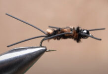
Once, for instance, Hervey-Jumper operated on a middle-aged man, an ophthalmologist and fly-fisher, who had developed a tumor above his left ear, in the brain area that controls the right hand. “I said, ‘Listen, we can remove this thing, but you probably won’t have full dexterity in your hand,’” Hervey-Jumper recalls telling the patient. “‘It’ll move, but it won’t do detailed work.’” Three months after the surgery, the patient emailed him a video. “He was tying a fly!” Hervey-Jumper relates. “I was shocked, because right around this time, I had another patient who had a tumor in the exact same location. It was nearly the same operation, same everything. One patient is fly-fishing and the other can’t even write.”
He wanted to understand why. “I thought there must be some underlying differences that cause some patients to recover to a fuller extent than others,” he says. He first tried looking for genetic factors, but after two years, he hit a dead end and decided to change tack.
By then, he had moved to UCSF, where he is an associate professor. Berger, who had trained Hervey-Jumper during his fellowship, had fought hard to recruit him. “I sensed that he was an extraordinary individual,” Berger says, “not just in terms of his technical skills but also in his ability to think outside the box.” Hervey-Jumper has cultivated this quality by surrounding himself with other out-of-the-box thinkers. Berger is one of them. Another is Michelle Monje, MD, PhD, a pediatric neuro-oncologist and neuroscientist at Stanford, whom he sought out as a mentor and collaborator.
Starting in the mid 2010s, Monje’s lab and others discovered that brain tumors called gliomas form a curious relationship with neurons in mice. Gliomas, which make up 80% of human brain cancers, arise from cells that are developing into glia, brain cells that manage neural connections. These lethal tumors don’t destroy neurons like bombs but instead invade them like weeds. Even more surprisingly, as the mouse studies revealed, glioma cells can cause neurons to fire more readily; in turn, this neural activity makes fast-growing tumors grow even faster.
Hervey-Jumper was intrigued. If neither genes nor gross anatomy could predict how a brain cancer patient would fare after surgery, he wondered, maybe something about their brain’s circuitry could. On Monje’s advice, he set about trying to replicate the mouse observations in his human patients. Using electrodes placed on the brain during surgery, he recorded neurons firing in the brains of glioma patients while they were awake and resting. Sure enough, cancerous brain regions were more active than healthy regions. But what did that mean for cognitive tasks like word-finding or fine motor movement?
“That’s where Eddie came in,” Hervey-Jumper says. Edward Chang, MD, a UCSF brain surgeon who did his residency under Berger, is now the Joan and Sanford Weill Chair of Neurological Surgery. Also a neuroscientist and the Jeanne Robertson Distinguished Professor of Psychiatry, Chang (who is not related to Susan Chang) has become the world’s leading expert in understanding how the brain generates speech. Researchers in his lab are so adept at capturing and deciphering patterns of activity in the brain’s language areas that they can decode entire words from these neural signals. They piggyback these experiments onto awake surgeries for epilepsy or cancer, discarding any signals from diseased brain tissue.
But those are precisely the signals that Hervey-Jumper was interested in. “Rather than throw out the tumor data, assuming it’s garbage,” he proposed, “let’s look at it specifically.”
With Eddie Chang’s guidance, Hervey-Jumper’s team began examining neural signals from patients like Gina who had gliomas in a speech area of their brain. What they found was astonishing. When a patient started to say a word, for instance, the tumorous area lit up in sync with the tissues around it, implying that neurons subsumed by cancer still behave somewhat like normal neurons. “That freaked me out,” Hervey-Jumper says, “because if you talk to these patients, their speech isn’t normal; they have a lot of impairments.”
Analyzing the speech signals more closely, however, the team saw that those from tumor-infiltrated neurons lacked intelligible information, like a staticky radio channel. Even Eddie Chang’s well-honed decoders couldn’t tell from the staticky signals whether patients were saying simple, monosyllabic words like “cat” or more complex, polysyllabic words like “aardvark.” Their tumors, it appeared, had rewired their brains like a careless electrician.
“These are incredibly exciting, mind-boggling discoveries,” Monje says. “They show that gliomas change not only the activity of neurons but also their connectivity; these tumors can remodel neurocircuitry in a way that is advantageous to the tumor but not necessarily to cognition.” She adds, “It’s very insidious.”
Since publishing his team’s initial findings in 2021 in Proceedings of the National Academy of Sciences, Hervey-Jumper launched the Glial Tumor Neuroscience Program to further explore the mechanisms involved. “For the longest time, people have thought about the brain and cancer as two separate things,” he says. “In fact, there are very important interactions that may give us clues about how to restore or preserve cognitive functions.”
As much as the prospect of death, he has learned, it’s brain cancer’s ability to hijack the mind that makes the diagnosis so devastating. “He tells me it’s one of the ugliest diseases he’s ever seen,” says Heather Hervey-Jumper, MD, a UCSF anesthesiologist and his wife of 21 years. “I think that’s why he likes the lab. You go into the operating room to face the beast, but the lab is where you find hope.”
Excising the tumor and his quest to preserve cognition
Removing the Monster
As the sedative slips into Gina’s vein, Hervey-Jumper starts in on the piece of tumor just below her brain’s surface, near the language areas. “I’m going to ask you silly questions so I can hear the fluency of your speech,” he tells her. He excavates as they talk, listening for trouble. He does not cut out the cancer but vacuums it up with a suction tool. Steadily, he sucks away the silky white flesh, taking care to avoid two large blood vessels snaking over it.
“Oooh, I felt that,” Gina says.
“I think the headache experience just spiked,” someone remarks. Local anesthesia can block sharp pain, but patients still feel a dull, mounting ache. Hervey-Jumper will soon need to let Gina sleep again. “People can’t just persist like that forever,” he says. “You only have a finite window.”
After he’s removed the first mass, he turns his attention to the dark underbelly of the cavity, where the rest of Gina’s tumor awaits. Here, the cancer sprawls around the corticospinal tract, a thick fan of neural fibers that connects the motor areas on the left side of the brain to the muscles on the right side of the body. Hervey-Jumper must navigate this precarious landscape a millimeter at a time, mapping the terrain as he tunnels through it.
With a stimulator – this time a device called a monopolar probe – he sends a beam of electricity into the wall of tumor before him. The strength of the current determines the distance it travels: 1 milliamp, for example, will travel 1 millimeter. Two neurophysiologists – John “J.P.” Clark, PhD, and Yanling Wang, MD – dial up the current until it strikes a bundle of motor fibers, causing the corresponding muscle to contract. Such movements are typically too small to see – sometimes a finger twitches or a lip curls – but they can be detected through electrodes stuck like postage stamps all over the patient’s body. “Threshold is 10 on lower leg,” Wang calls out, and Hervey-Jumper knows he is 10 millimeters – about the width of a paper clip – from the neurons that control Gina’s leg.
Berger once told him that doing awake surgery is like flying a plane at night: You have to trust your brain-mapping data the way a pilot trusts their aircraft’s radar system. He sucks up a speck of tumor and stimulates again. “Now 8.5,” Wang says.
They continue like this as Hervey-Jumper vacuums deeper and deeper, closer and closer to the corticospinal tract. Then he stops. He is 5 millimeters away.
“Gina, can you wiggle your toes for me?” he says. “Can you point your toes toward your nose? Good,” he concludes. “I don’t want to go too much further or you’re going to have a drop foot. If you need to run, you’ll still be able to run.” But, he continues, “let’s see what else we can get.”
He picks his way cautiously along the tumor’s margins, shaving off another millimeter here, another there.
“Okay, we’re done,” he says at last. “We shouldn’t go any further.”
Gina drifts out of consciousness. In the following days, as her brain swells from the surgery, she will have some trouble walking and will struggle to find the right words, but these impairments will be only temporary. Hervey-Jumper replaces her dura, bone, and scalp and leaves Choy, the resident, to sew the last few stitches. His next patient is waiting for him.
The early experiences that shaped a surgeon

Credit: Source link


